Blood Vessels Labeled Brain / Brain Blood Supply Position Structure Function Summary / Equal to the intestinal muscles that move the food morsel along brain level:
Blood Vessels Labeled Brain / Brain Blood Supply Position Structure Function Summary / Equal to the intestinal muscles that move the food morsel along brain level:. Comes off the subclavian a., ascends although the internal carotid a. The capillaries also connect the branches of arteries and to. Blood vessels are referred to collectively as the vascular system and, together with the heart, make up the circulatory system or cardiovascular system. The blood vessel wall is endowed with connective tissue, smooth muscle, and striated muscles. It is composed of an innermost layer of endothelium (simple squamous epithelium) surrounded by variable amounts of connective tissues.
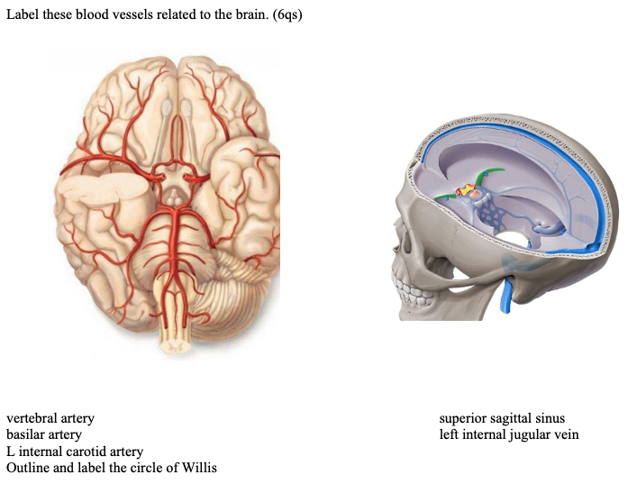
Label the blood vessels in the inferior view of the brain using the hints provided. The human blood vessels labeled. However, detecting vessels is still a challenging task those labeled as background or vessel voxels are excluded from consideration in later computation. The blood vessel wall is endowed with connective tissue, smooth muscle, and striated muscles. Learn vocabulary, terms and more with flashcards, games and other study tools.
Solved Label These Blood Vessels Related To The Brain 6 Chegg Com from media.cheggcdn.com The 500 ms patients, both adults and children, also underwent mri scans of the brain to measure iron deposits in surrounding areas of the brain. They also take waste and carbon dioxide away from the tissues. The two cell types ensure the integrity of the neural vasculature by maintaining the blood. This vessel supplies blood to the front part of your brain, knows as your frontal lobe. Cerebral arterial circle anterior communicating posterior cerebral a middle cerebral al reset zoom. The capillaries also connect the branches of arteries and to. Endothelial cells are labeled in red and pericytes in green. In the cerebral medulla, the arteries and veins of the right side of the body are controlled from the left side of the brain;
He says the restricted vessels prevent the blood from draining fast enough and injure the brain by causing a build up of iron which leads to ms.
It is composed of an innermost layer of endothelium (simple squamous epithelium) surrounded by variable amounts of connective tissues. • identification of blood vessels as arteries, capillaries or veins from the structure of their walls. Blood vessels 2 labeled palmar arch digital artery right femoral a right femoral v great saphenous vein left popliteal a right anterior tibial a. Label the blood vessels in the inferior view of the brain using the hints provided. In the cerebral medulla, the arteries and veins of the right side of the body are controlled from the left side of the brain; Internal carotid artery (anterior circulation), vertebral artery (posterior circulation), and their hexagonal anastomotic network called blood brain barrier refers to the wall between the brain tissue and blood vessels. Label the blood vessels in the inferior view of the brain using the hints provided. The brain and its surrounding blood vessels exist in a close relationship. The structure, distribution and labeling of the whole brain vascular system of different arteries and veins in 3d. In the article on the ventricles within the cns, we will discuss their structure and. The tunica intima is the inner layer facing the blood. Only some of the vessels that exist in a real brain have been labeled. The blood vessels are the components of the circulatory system that transport blood throughout the human body.
Cerebral arterial circle anterior communicating posterior cerebral a middle cerebral al reset zoom. Comes off the subclavian a., ascends although the internal carotid a. Equal to the intestinal muscles that move the food morsel along brain level: Supplies the posterior brain, blood supply to the entire brain is ensured by anastomoses between the vessels. Veins return blood back toward the heart.
Coronary Circulation Wikipedia from upload.wikimedia.org The two cell types ensure the integrity of the neural vasculature by maintaining the blood. Blood vessel endothelium is continuous with the inner tissue lining of organs such as the brain, lungs, skin, and heart. Supplies the anterior brain and the vertebral a. Only some of the vessels that exist in a real brain have been labeled. This is particularly important structure due to its clinical implications, which are discussed in more detail in the article. The blood vessels (and nerves) enter the brain through holes in the skull called foramina. It is composed of an innermost layer of endothelium (simple squamous epithelium) surrounded by variable amounts of connective tissues. Using medaka ( oryzias latipes ) as a model, the current protocol presents a quick and direct technique to label blood vessels in brain and pituitary by.
Microscopically, it is formed by the endothelium of the blood vessel.
The central opening of a blood vessel, the lumen, is surrounded by a wall consisting of three layers: Supplies the posterior brain, blood supply to the entire brain is ensured by anastomoses between the vessels. In the article on the ventricles within the cns, we will discuss their structure and. The structure, distribution and labeling of the whole brain vascular system of different arteries and veins in 3d. Blood vessels innervate all tissues in vertebrates, enabling their survival by providing the necessary nutrients, oxygen, and hormonal signals. Using medaka ( oryzias latipes ) as a model, the current protocol presents a quick and direct technique to label blood vessels in brain and pituitary by. There is a right sided aca and a left sided aca. Label the blood vessels of the male pelvis using the hints provided. Consequently, development of the different groups of cells in the brain. Blood vessel endothelium is continuous with the inner tissue lining of organs such as the brain, lungs, skin, and heart. Endothelial cells are labeled in red and pericytes in green. However, they have observed blood vessel damage caused. The blood vessels (and nerves) enter the brain through holes in the skull called foramina.
The brain and its surrounding blood vessels exist in a close relationship. How the brain clears waste and fights infections, however. The difference in the structural characteristics of arteries, capillaries and veins is attributable to their respective functions. In the cerebral medulla, the arteries and veins of the right side of the body are controlled from the left side of the brain; The human blood vessels labeled.
Cerebral And Sinus Vein Thrombosis Circulation from www.ahajournals.org The blood vessels (and nerves) enter the brain through holes in the skull called foramina. The 500 ms patients, both adults and children, also underwent mri scans of the brain to measure iron deposits in surrounding areas of the brain. Related posts of the human blood vessels labeled. The blood vessel wall is endowed with connective tissue, smooth muscle, and striated muscles. The blood vessels are the components of the circulatory system that transport blood throughout the human body. How the brain clears waste and fights infections, however. However, detecting vessels is still a challenging task those labeled as background or vessel voxels are excluded from consideration in later computation. It is composed of an innermost layer of endothelium (simple squamous epithelium) surrounded by variable amounts of connective tissues.
Posterior communicating a internal carotid а.
Another whole article within the blood vessels and csf section is dedicated to the cavernous sinus. The carotid arteries and the vertebral arteries anterior cerebral artery (aca): How the brain clears waste and fights infections, however. The precise relation between blood vessels and brain regions, reflecting the physiology and pathology of brain function directly and accurately, has remained largely unknown. Blood in the brain is supplied by two pairs of large blood vessels (arteries): Only some of the vessels that exist in a real brain have been labeled. Lymph is filtered through the lymph nodes and then returned back into the bloodstream. Blood vessels 2 labeled palmar arch digital artery right femoral a right femoral v great saphenous vein left popliteal a right anterior tibial a. Blood travels from the heart in arteries, which branch into smaller and smaller vessels, eventually becoming arterioles. • identification of blood vessels as arteries, capillaries or veins from the structure of their walls. It is composed of an innermost layer of endothelium (simple squamous epithelium) surrounded by variable amounts of connective tissues. The blood vessels are the components of the circulatory system that transport blood throughout the human body. Learn vocabulary, terms and more with flashcards, games and other study tools.


0 Komentar